
For patients diagnosed with end-stage renal disease (ESRD) requiring chronic hemodialysis, establishing a dependable, durable vascular access is literally a lifeline. Among available hemodialysis modalities, the Arteriovenous (AV) Fistula remains the universally acknowledged “gold standard” worldwide, supported by international nephrology and vascular surgery guidelines.
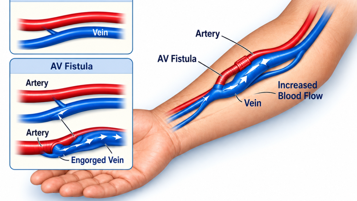
An AV fistula is created surgically by directly joining a patient’s native artery to an adjacent vein, typically at the wrist (radiocephalic) or inside the elbow crease (brachiocephalic). Under normal physiologic conditions, veins have thin walls and low-pressure blood flow, making them incapable of sustaining repeated punctures with large dialysis needles or delivering the high flow rates (typically 300 to 500 mL/min) required by a hemodialysis machine. When surgically anastomosed to an artery, high-pressure arterial blood rushes into the vein. Over several weeks or months, the vein undergoes a process known as “maturation”—thickening its muscular walls and dilating significantly so it can withstand multiple weekly needle cannulations.
Compared to synthetic arteriovenous grafts (AVG) or Central Venous Catheters (CVC), native AV fistulas provide superior clinical advantages:
- Lower Infection Rates: Because the fistula utilizes the patient’s own native vessels, it exhibits the lowest incidence of severe bacteremia and systemic sepsis compared to foreign synthetic materials or plastic catheters.
- Long-Term Patency: Native fistulas last significantly longer than grafts, often functioning reliably for years or even decades with proper surveillance.
- Fewer Thrombotic Complications: Fistulas are far less likely to develop sudden blood clots (thrombosis) compared to prosthetic conduits.
- Superior Blood Flow: Generates optimal, stable blood flow rates that ensure high-efficiency dialysis clearance during every treatment cycle.
Successful fistula preservation relies heavily on regular diagnostic surveillance. Over time, recurring needle punctures and hemodynamic shear stress can cause neointimal hyperplasia, leading to progressive stenosis (narrowing) near the anastomosis or outflow veins. Vascular specialists utilize high-resolution color Doppler ultrasound to monitor flow velocity, vessel diameter, and structural patency.
When narrowing occurs, minimally invasive endovascular balloon angioplasty (fistuloplasty) can be performed without open surgical incisions, gently dilating the narrowed segment to maintain adequate dialysis access. Patients with advancing kidney disease should seek early vascular consultation for vessel mapping well before beginning dialysis, ensuring adequate time for the fistula to mature naturally and safely.